
Transmission Electron Microscopes, or TEM:
The TEM is when electrons pass through a very thin sample there is contrast in the denser parts, because the electrons don't pass through as easily. A 2D image is produced. The largest magnification possible with the TEM is x500,000.

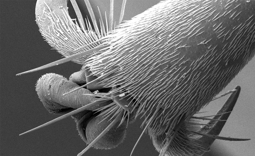
The Scanning Electron Micrscope, or SEM:
The electron beam is fired at the sample, but doesn't pass through - they're reflected away to give a 3D image. The highest magnification with the SEM is around x100,000.
Summary:
The Electron Microscope has a much higher resolution than a ligh microscope (0.1nm!). We can also use the TEM to view the organelles inside cells, rather than the top layer of the specimen. The SEM can provide a unique 3D image when required. Some of the troubles involved in using an Electron Microscope are that the microscope is a difficult piece of equipment to master, and that the microscope must have a vaccuum (so the Electrons don't rebound off air particles etc) - greatly increasing the cost of the microscope.
No comments:
Post a Comment